
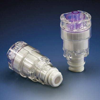
Q-PLUS Labs, the West Coast Partner of Bruker alicona, Introduces the µCMM, the World’s First Purely Optical CMM, into Service
After months of extensive research followed by planning, preparations, installation, calibration, training, qualification, and validation, we are proud to announce the deployment of the Bruker alicona µCMM, an extraordinary addition to our vast array of dimensional metrology technology and capability. The µCMM is the first purely optical CMM machine on the market and the The team at Q-PLUS Labs is very excited to be the first independent accredited and multi-certified* dimensional metrology lab in the world to put it into service. The µCMM is used to measure extremely tight tolerances with ultra-high accuracy. Users combine advantages from tactile coordinate measuring technology and optical surface measuring technology and measure the dimension, position, shape and roughness of components with only one extraordinary sensor. *(as defined by our ISO 17025 accreditation, ISO 9001 registration, AS9100 & ISO 13485 certifications)
The optical CMM offers high geometric accuracy of several optical 3D measurements in relation to each other, enabling the measurement of small surface details on large components and precisely determining the position of these individual measurements in relation to each other. The spectrum of measurable surfaces includes all common industrial materials and composites such as plastics, PCD, CFRP, ceramics, chrome, silicon. Simple operation is implemented by single-button solutions, automation and ergonomic control elements such as a specially designed controller. Air-bearing axes with linear drive enable wear-free use and highly accurate, fast measurements.
|
|
|---|
THE µCMM OFFERS
The spectrum of measurable surfaces is largely material-independent and includes all materials and composites commonly used in the industry, from matte to polished or mirrored components.
Alicona measurement systems offer the functionalities of a surface-roughness measuring device as well as a coordinate measurement system combined into one system. As a result, quality assurance becomes more cost effective, is easier to execute and delivers results more efficiently.
All components, including the moving axes, operate contact-free. Air-bearing linear drive axes enable wear-free operation and high-precision, fast measurement. This makes μCMM ideal for permanent use in production.
Below are Some Examples of the µCMM in Use
Images speak volumes to the machine’s capabilities. We’ve presented some examples below of some of the most difficult features to measure in dimensional metrology today. While Bruker alicona has very informative brochures and data sheets with remarkable images, we wanted to show off some applications we selected ourselves and performed here right on our own μCMM.
Example #1 – Scan of the Sharp Edge of a Razor Blade
A seemingly simple razor blade is representative of geometry that is notoriously difficult, almost impossible to measure completely and properly using other equipment and technology whether contact or non-contact. The challenge for contact stylus devices comes from the fact that the stylus, a kind of blade itself with two surfaces acutely angled to each other, can only measure with the very small radiused tip on the stylus so as the stylus tries to go over the sharp tip of the razor blade, it will “shank out” on one of the angled surfaces, preventing full measurement of the razor blade tip geometry. One has to tilt the razor blade toward the stylus to get one part of the geometry, then reposition, tilting the razor blade away from the stylus to get the rest of the geometry. However, the shared trace data or overlap of the two traces is very difficult, if not impossible, to align properly for subsequent integration of the combined trace to obtain the incredibly small razor blade tip radius and the angle of the adjoining surfaces.
It is similarly difficult for other non-contact sensors because none of them can handle very high slope (nearly vertical surfaces), that again requires performing the measurement in segments and trying to piece together the trace data. The μCMM addresses this problem not just in one but two ways:
-
- The Vertical Focus Probing can measure flanks of 90 degrees or higher! The sensor has a wide detection cone that allows the vertical objective to effectively see sideways, an impossibility for all conventional vision systems or any other visible light non-contact sensor. Lateral probing of vertical surfaces was limited to tactile measuring systems until now.
- Not only does the μCMM have a true 4th axis—itself a rarity, it also has a true 5th axis. This allows the part to be articulated in any direction, allowing for a full complete measurement without the need to manually piece together traces or scans (or try to, if even possible). Repeat, these are NOT mere rotary indexers where the original coordinate system is lost, but axes that preserve it!
Example #1 – Razor Blade – Photograph With Area of Interest Circled in Red

Example #1 – Razor Blade – Post 3D Scan – Magnified View Showing 2D Cross Section

The apparent surfaces are actually a triangulated mesh of tens of thousands of 3D measurement points. A planar cross section is taken perpendicular to the axis of the sharp edge.
Example #1 – Razor Blade, 2D Cross Section – Close Up Planar View

The small gray areas in the left image are imperfections in the blade surface behind the cross section. Note the incredibly small 0.0003” radius at the tip showing that even a brand new razor blade is not actually perfectly sharp.
Example #2 – Hypodermic Needle – Scan to Observe Sharpness of the Tip
In our next example, we measure the tip of a hypodermic needle, also a notoriously difficult measurement task. Since such small geometry is virtually impossible to measure using conventional contact probing, high magnification vision systems can provide close up views and even some 2D measurements of edges. However, the actual needle tip, always has degrees of less-than-perfect geometry including very small burrs and surface distortions that can be important to characterize.
However, the μCMM is not subject to any of these limitations. It’s sensor is not 2D like a vision system camera, nor is it limited by how small of a contact probe or stylus would be necessary to even attempt such measurements using contact. And conventional 3D scanners stand no chance of achieving the resolution and accuracy needed to truly capture the nature of the needle tip. Even other 3D noncontact sensors such as white light interferometry or confocal laser microscopy, while able to capture some of the tip geometry, simply aren’t typically integrated into a whole system that can capture ALL of the geometry in one run.
Example #2 – Hypodermic Needle – Photograph With Area of Interest Circled in Red

Example #2 – Hypodermic Needle – High Magnification Photograph Further Showing Very Small Area of Interest Circled in Red

The tiny burr and surface distortions can already be seen but are not yet 3D scanned.
Example #2 – Hypodermic Needle – Post 3D Scan Close Up Characterizing the Nature, Shape, and Size of the Tip

What might appear to be a 3D CAD model of the actual needle tip is actually a triangulated mesh of hundreds of thousands of measured 3D coordinates.
Example #3 – Laser-Drilled Ø0.0035” Hole in a Catheter Tip – Scan to Measure Size, Form AND Negative Taper
In this example, there are not one but multiple measurement challenges: measure the hole size and position on a small cylindrical and spherical tip where due to the nature of manufacture, the holes get bigger as they go deeper.
For starters, there are no typical CMM probe styli made this small, and if there were, they’d be extraordinarily fragile and break very easily. While it may seem just a matter of having a high enough magnification lens and intense enough lighting on a vision system to measure incredibly small holes such as those shown here, a vision systems need to measure edges, something they can focus on to produce readings. With holes this small, the clarity of the circular edge becomes critically important, but as we have shown in the razor blade and the hypodermic needle examples above, the edges are never perfectly sharp. This presents limitations (sometimes severe) for a conventional vision system. Even fiber optic probe accessories, another expensive and fragile tool, it is difficult, if not impossible, to find at this size. And even if there was a small enough fiber optic probe, it would have to be attached to a 5-axis machine to obtain all the measurements here and would be very slow.
How about micro CT scanning? We have one in our lab, it is a solid option for many applications, but this part is made of a heavy metal and not very conducive to such an approach. CT scanning, while certainly essential to a modern dimensional metrology lab, still involves many steps from imaging in dozens if not hundreds of directions with sufficient energy, to a voxel file, to a point cloud, to post-processing of the point cloud, to measurement results. And that’s only if it is accurate enough. It’s hard to get that ultra-high resolution and accuracy at the high levels of X-ray energy needed to see through such a dense part, even if the part is small.
The μCMM offers a superior approach, where a pre-programmed part can be loaded, and the measurements quickly performed ONLY where they are needed (instead of the entire part). This way, the measurement data is automatically processed, and the results are output in minutes.
What about the holes getting bigger as they go deeper? Surely that would limit any optical system, right? Wrong. Because of the nature of the μCMM Focus-Variation sensor, it can not only measure the part of the hole at the surface but measure the cylindrical walls of this tiny hole even as it increases in diameter with depth!
Example #3 – Photograph of Laser-Drilled Ø0.0035” Holes in a ¼” Diameter Cylindrical Catheter Tip

Example #3 – Post Scan and Measurement of Laser-Drilled Ø0.0035” Hole – Highly Magnified View

The apparent cylindrical surfaces are actually a triangulated mesh of tens of thousands of 3D measurement points. In the left image, a cross-section is taken perpendicular to the axis of the tapered cylindrical hole (essentially a slight conical). In the right image, a circle is fit using a least-squares algorithms to determine diameter size and roundness at a given depth. A cylinder or conical element could also be fit through the measurement data to measure, in this case, negative taper where the diameter increases with depth.
Example #4 – Plastic Injection Molded Energy Director – Scan for Contour Definition
Another challenging measurement is the geometry of energy directors commonly found in plastic injection molded parts that become fused into an assembly. A complex process using ultrasound to fuse or “weld” precision plastic parts together requires that relatively simple but highly accurate and consistent geometry exists at or near the periphery of the given part. The energy director is typically a triangular cross-section with the base at the bottom and the tip at the top. This geometry can appear in hard-to-reach areas, such as the example shown below. The size of the energy director is incredibly small, often less than 0.015” while also having tight tolerances, making it impractical to measure using a CMM. A precision contour tracer with a nearly sharp stylus is one way to measure this geometry, but measuring the features at multiple locations requires reorienting the part over and over and can be time-consuming since careful alignment is required.
The μCMM can quickly and even more accurately perform these measurements. Since the sensor gathers 3D “patches” of measurement data containing thousands to tens of thousands of points or more, it can capture geometry around the entire part allowing for an unlimited number of cross sections or even 3D analyses of actual-to-nominal geometry.
Example #4 – Photograph of Plastic Injection Molded Energy Director

Example #4 – Plastic Injection Molded Energy Director – Post Scan
Again, the triangulated point cloud above visualizes the massive measurement data just obtained.
Example #4 – Plastic Injection Molded Energy Director – Post Scan, Cross Section, and Measurement

In the left image, a cross section is taken perpendicular to the path of the triangle-shaped energy director. In the right image, the measurements are extracted from the cross-section geometry producing readings for the angle, height, and tip radius (total size less than .020”).
Example #5 – 100 Grit Sandpaper – High Resolution 3D Scan and Visual Depiction
Lastly, just for fun, we included an ultra-high resolution 3D scan of something most everyone is familiar with: sandpaper, in this case 100 grit. On the CAMI scale, 100 grit, which equates to a grain size of 141 microns (.0055”), is very small.
Example #5 – Photograph of 100 Grit Sandpaper and the Subsequent 3D Nano Scan of a Small Area

Have a Project in Mind? Get in Touch with Us for More Information.
We hope you found these examples to be helpful, informative and interesting. See what the Brucker alicona μCMM can do for you by sending us your application. Our μCMM is available for dimensional measurement and inspection services and is already being utilized on several applications. It is also available to run applications for customers who might be interested in acquiring their own μCMM. We will also be hosting a workshop on the μCMM here at Q-PLUS Labs very soon!
Contact us today to learn more. We would be glad to answer any questions about the µCMM and Q-PLUS Labs’ Optical CMM services.


Leave A Comment
You must be logged in to post a comment.